

Spinal stenosis is a narrowing of the gaps in the spine. It puts pressure on the nerves that run through the spine. Most commonly, spinal stenosis occurs in the neck and lower back. Changes with age in the spine are a common cause of spinal stenosis.
Some people with spinal stenosis don’t experience any signs or symptoms. While others may have tingling, numbness, and weakness in their arms and legs, along with back and neck pain. When walking up or down a ramp, a slope, or steps, pain may strike more quickly. It is usually eased by sitting or leaning over. In this blog, I will tell you the types, causes, symptoms, prevention, and diagnosis of spinal stenosis.
Types of Spinal Stenosis
Spinal stenosis is categorised based on where the problem develops in the spine. It is possible to have multiple types. There are two types of spinal stenosis:
Cervical stenosis: The narrowing occurs in the region of the spine in the neck.
Lumbar stenosis: It is the most common type of spinal stenosis. The narrowing occurs in the lower back region of the spine.
Common Causes of Spinal Stenosis
One or more of the following reasons commonly induce spinal stenosis:
Spinal osteoarthritis.
When the smooth cartilage covering the facet joints, which link the backs of adjacent vertebrae, begins to break down, bones rub against each other, which can lead to the production of osteophytes or bone spurs. The resultant osteophyte growth and inflammation may cause the foramina to narrow.
Degenerative disc disease.
The intervertebral foramina narrows as the discs lose moisture and begin to flatten. A bulging disc can also push into the spinal canal. Disc degeneration can also increase the pressure on the facet joints, speeding up their degeneration.
Ligament thickening or buckling.
Ligaments in the spinal canal can ossify (harden and become bony tissue) and intrude on the spinal cord or neighbouring spinal nerves. As spinal degeneration proceeds, some ligaments may become more prone to buckling into the spinal canal.
Other conditions, such as spinal deformities or cyst growths, may also lead to spinal stenosis.
Symptoms Of Spinal Stenosis
Many people with spinal stenosis have signs of it on an MRI or CT scan but no symptoms. When symptoms do happen, they usually start slowly and get worse over time. The symptoms vary depending on where the stenosis is located and which nerves are affected.
Symptoms of the Cervical Spine
- Weakness in an arm, hand, foot, or leg
- Neck pain
- Tingling or numbness in an arm, hand, foot, or leg
- Walking and balance problems
- In extreme cases, bladder or bowel dysfunction (urinary urgency and incontinence)
Symptoms of the Lumbar Spine
- Back pain
- Weakness in a leg or foot
- Tingling or numbness in the leg or foot
- A pain or cramping in one or both legs that occurs when you stand for a long period of time or walk, which usually goes away when you bend forward or sit.
When Spinal Stenosis Is Serious?
When spinal stenosis gets severe, neurological deficits such as myelopathy, radiculopathy, and/or cauda equina syndrome might emerge. Permanent numbness and/or paralysis can occur if a spinal nerve or the spinal cord is compressed for a lengthy period of time. Any weakness or numbness that radiates into the arm(s) or leg(s), as well as any issues with coordination or bowel/bladder control, should be treated as soon as possible.
What Are The Methods To Prevent Spinal Stenosis?
You can’t completely prevent spinal stenosis because the majority of the causes are age-related “wear and tear” disorders like osteoarthritis and loss of bone and muscle mass. You can, however, take steps to reduce your risk or slow the growth of the disease, such as:
- Maintain a healthy diet and a healthy body weight.
- Please don’t smoke. If you smoke, give it up. To quit, seek help from your healthcare practitioner.
- Maintain a healthy posture.
- Keep active and avoid painful exercises. Before beginning a home fitness regimen, consult your doctor or physical therapist. Avoid too much sleep, as it can be dangerous rather than beneficial.
Diagnosis Of Spinal Stenosis
Your medical history will be reviewed, questions regarding your symptoms will be asked, and a physical exam will be performed. The doctor may feel your spine during your physical exam, pressing on different areas to discover the area of pain. Your doctor may probably ask you to bend in different directions to determine if different postures of your spine cause pain or other symptoms. He may also assess your movement, balance, and walking patterns, as well as your arm and leg strength. Imaging tests will be performed on your spine to determine the main location and extent of the disease. Imaging studies may involve the following:
X-rays
X-rays emit a limited quantity of radiation and can reveal changes in the bone structure such as disc height reduction and the formation of bone spurs that narrow the spine’s space.
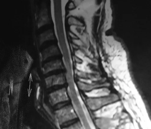
MRI
Magnetic resonance imaging (MRI) creates cross-sectional images of the spine using radio waves and a strong magnet. MRI pictures show the discs, nerves, spinal cord, and the existence of any tumours in great detail.
CT or CT myelogram.
A computed tomography (CT) scan uses a series of X-rays to produce cross-sectional images of the spine. A contrast dye is used in a CT myelogram to help show the spinal cord and nerves more clearly.
Click here to contact for more information